EMU Instruments for Scanning Electron Microscopy

MFZ Building 1st floor EMU Room 1.039 Scanning Electron Microscope (SEM) ZEISS Crossbeam 540 FIB
Cryo-SEM with capella column with Ga+++ FIB, in-lens, SE2- and energy selective detectors, STEM, GIS, resolution 0.7 nm at 30KV with Atlas software package
for
- standard scanning electron microscopy
- focused ion beam investigations --> 3D reconstructions
- cryo scanning electron microscopy

MFZ Building 1st floor EMU Room 1.039 Cryo Scanning Electron Microscopy Preparation System Quorum PP3010T
Preparation table + preparation chamber (Quorum PP3010T) which is directly flunshed onto the SEM
for
- cryo scanning electron microscopy
- freeze etching and fracturing scanning electron microscopy

MFZ Building 1st floor EMU Room 1.035 Critical Point Dryer Leica EM CPD 300
With this device scanning electron microscopic samples are critical-point-dried which is necessary before sputtering

MFZ Building 1st floor EMU Room 1.037 Sputter-Coater Leica ACE 600
With this device the surfaces of specimens are sputter coated with metals (platinum, palladium, gold or chrome) for better visualisation.

MFZ Building 1st floor EMU Room 1.039 Sputter-Coater Quorum Q 150TES
For some samples it is reasonable to perform carbon sputter coating before investigation on the scanning electron microscope. This device exclusively is used for that purpose.

MFZ Building 1st floor EMU Room 1.037 Laboratory microwave Bio Wave Pro Plus
Lab microwave with magnetic stirrer and vaccum system for
- quick embedding of electron microscopic samples
- treatment of tissue before paraffin- or immune fluorescence embedding
- decalcification (e.g., of bones)

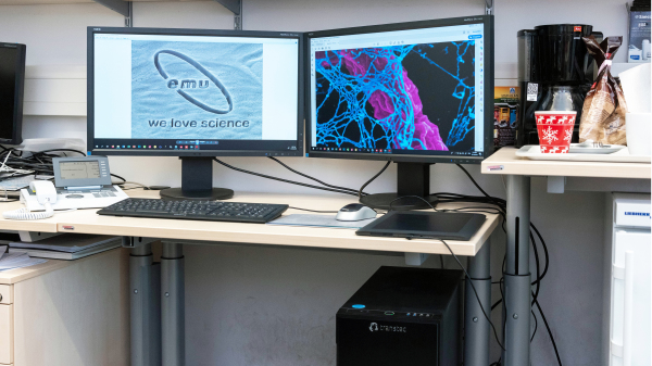
MFZ Building 2nd floor EMU Room 2.013 Workstations for image processing & reconstructions
High-end workstations for professional image processing & segmentation
for
- working with + segmentation of large image stacks
- three-dimensional visualisation of segmented structures

